THE NUZZO LETTER IN THE NEWS
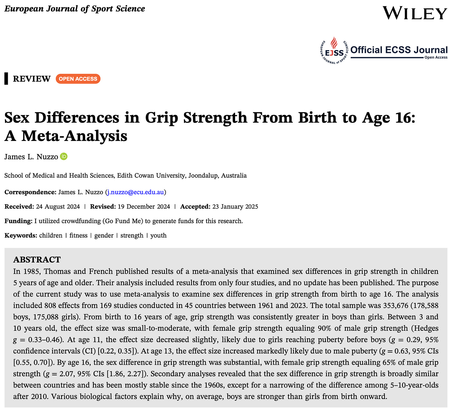
Sex Differences in Grip Strength From Birth to Age 16: A Meta‐Analysis
European Journal of Sport Science
Abstract: In 1985, Thomas and French published results of a meta-analysis that examined sex differences in grip strength in children 5 years of age and older. Their analysis included results from only four studies, and no update has been published. The purpose of the current study was to use meta-analysis to examine sex differences in grip strength from birth to age 16. The analysis included 808 effects from 169 studies conducted in 45 countries between 1961 and 2023. The total sample was 353,676 (178,588 boys, 175,088 girls). From birth to 16 years of age, grip strength was consistently greater in boys than girls. Between 3 and 10 years old, the effect size was small-to-moderate, with female grip strength equaling 90% of male grip strength (Hedges g = 0.33–0.46). At age 11, the effect size decreased slightly, likely due to girls reaching puberty before boys (g = 0.29, 95% confidence intervals (CI) [0.22, 0.35]). At age 13, the effect size increased markedly likely due to male puberty (g = 0.63, 95% CIs [0.55, 0.70]). By age 16, the sex difference in grip strength was substantial, with female grip strength equaling 65% of male grip strength (g = 2.07, 95% CIs [1.86, 2.27]). Secondary analyses revealed that the sex difference in grip strength is broadly similar between countries and has been mostly stable since the 1960s, except for a narrowing of the difference among 5–10-year-olds after 2010. Various biological factors explain why, on average, boys are stronger than girls from birth onward.
(Nuzzo note: I would like to express my gratitude to the subset of subscribers who supported this research with donations made at the designated Go Fund Me page and the DonorBox at The Nuzzo Letter. Thank you!)
PODCASTS AND PRESENTATIONS
Australia’s $4.7 BILLION Aid Scandal – You Won’t Believe Where It’s Going!
Dave Right Now
ARTICLES AND ESSAYS
Argentinian President Calls Out Pervasive Dishonesty and Hypocrisy of Feminism
Domestic Abuse and Violence International Alliance (DAVIA)
Where does the British public stand on transgender rights in 2024/25?
YouGov UK
13 Steps to Make America Male Friendly Again
Men are Good
NFL Fumbles Super Bowl Opportunity
In His Words
New book exposes details on Claudine Gay Harvard plagiarism scandal
The College Fix
University of Alabama philosophy class studies ‘oppression’ of ‘sizism’
The College Fix
Professors call for tax on meat to address ‘climate change’
The College Fix
Journal of Science in Sport and Exercise
Abstract: Purpose - This study compared absolute and relative measurements of peak torque (PT), mean power (MP), and rate of velocity development (RVD) during isometric and isokinetic elbow flexion muscle actions in pre- and post-pubescent males and females. Methods - Forty children participated (pre-pubescent: mean ± 95% confidence interval age = 9.79 ± 0.35 years, n = 10 males, n = 10 females; post-pubescent: 17.23 ± 0.58 years, n = 10 males, n = 10 females). Ultrasound images quantified muscle cross-sectional area (CSA) of the elbow flexors. Participants completed maximal elbow flexion muscle actions at 0, 60, 120, 180, 240, and 300°/s. Results - As expected, the post-pubescent group achieved greater absolute measures of PT, MP, and RVD at all velocities (P ≤ 0.015). When normalized to CSA and strength, differences in PT were mostly eliminated (P ≥ 0.136), but differences in MP and RVD remained (P ≤ 0.012). Conclusion - The post-pubescent groups maintained greater MP and RVD at each movement velocity, suggesting that muscle size and strength cannot fully account for age-related differences in time-dependent measurements of muscle performance, such as power and acceleration. Instead, these performance indicators may be related to neuromuscular adaptations during normal growth, development, and biological maturation processes, such as augmentations in motor unit recruitment strategies and increases in action potential conduction velocities.
Human lower leg muscles grow asynchronously
Journal of Anatomy
Abstract: Muscle volume must increase substantially during childhood growth to generate the power required to propel the growing body. One unresolved but fundamental question about childhood muscle growth is whether muscles grow at equal rates; that is, if muscles grow in synchrony with each other. In this study, we used magnetic resonance imaging (MRI) and advances in artificial intelligence methods (deep learning) for medical image segmentation to investigate whether human lower leg muscles grow in synchrony. Muscle volumes were measured in 10 lower leg muscles in 208 typically developing children (eight infants aged less than 3 months and 200 children aged 5 to 15 years). We tested the hypothesis that human lower leg muscles grow synchronously by investigating whether the volume of individual lower leg muscles, expressed as a proportion of total lower leg muscle volume, remains constant with age. There were substantial age-related changes in the relative volume of most muscles in both boys and girls (p < 0.001). This was most evident between birth and five years of age but was still evident after five years. The medial gastrocnemius and soleus muscles, the largest muscles in infancy, grew faster than other muscles in the first five years. The findings demonstrate that muscles in the human lower leg grow asynchronously. This finding may assist early detection of atypical growth and allow targeted muscle-specific interventions to improve the quality of life, particularly for children with neuromotor conditions such as cerebral palsy.
HISTORICAL ARTICLES AND ESSAYS
Sex differences in the activity level of infants
Infant and Child Development, 1999
Abstract: A gender difference in motor activity level (AL) is well established for children, but questions about the existence and nature of an infant sex difference remain. To assess these questions, we applied meta-analytic procedures to summarize 46 infancy studies comprising 78 male–female motor activity comparisons. Our results showed that, as with children, male infants were more active than females. Objective measures of infant AL estimated the size of this difference to be 0.2 standard deviations, though subjective parent-report measures estimated the difference to be smaller. We argue that this early sex difference in activity level is biologically based. However, socialization processes, such as gender-differentiated expectations and experiences, in conjunction with further sex-differentiated biological developments, amplify this early difference to produce the larger gender differences in activity found during childhood.
RUBBISH BIN
No rubbish this week!
SUPPORT THE NUZZO LETTER
If you appreciated this content, please consider supporting The Nuzzo Letter with a one-time or recurring donation. Your support is greatly appreciated. It helps me to continue to work on independent research projects and fight for my evidence-based discourse. To donate, click the DonorBox logo. In two simple steps, you can donate using ApplePay, PayPal, or another service. Thank you!
If you prefer to donate to a specific project, please see the Go Fund Me page for my current research on sex differences in muscle strength in children.


